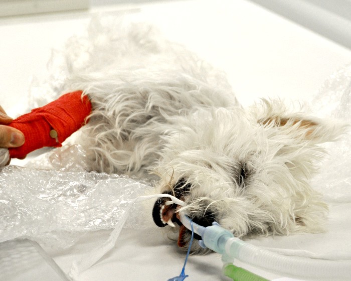
This view is particularly useful for demonstrating small pneumothoraces, emphysema or loculated pleural fluid.
The animal is placed in lateral recumbency. To demonstrate trapped pleural fluid, the affected side should be uppermost; to demonstrate emphysema, the affected side is placed down.
The x-ray cassette is positioned perpendicular to the spine on the table with a block – for example, a foam wedge or sandbag behind the cassette – to stop it from tipping backwards. The x-ray tube head is rotated so that it is parallel with the table and the x-ray beam is horizontal.
The x-ray beam should be directed towards an external wall and care should be taken that no people are on the other side of the wall.

Leave a Reply