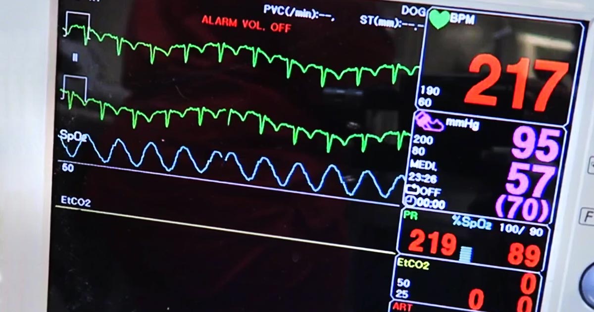
Pulse oximetry is a useful, non-invasive method of measuring a patient’s oxygen saturation (SO2) and, under normal physiological circumstances, correlates well to the arterial oxygen saturation (SaO2).
However, despite its ease of use and accessibility, it is not infallible. Circumstances exist that will undermine the accuracy of these readings – some with dire consequences if not recognised.
Others causes are more technically associated, but also needs recognition.
Unequal to task
Pulse oximetry is incapable of assessing:
- a patient’s haemoglobin levels
- the haemoglobin’s functionality
- the patient’s partial pressure of arterial carbon dioxide (PaCO2)
The former is particularly apparent in anaemic patients, where peripheral capillary oxygen saturation (SpO2) readings could be greater than 95%, but animals still severely hypoxic. This is because the total numbers of haemoglobin is reduced; therefore, overall oxygen-carrying capacity is also decreased.
Similarly, haemoglobin can be fully saturated with carboxyhaemoglobin or methaemoglobin strands, giving a misleadingly high SpO2 reading, yet patients are severely oxygen deprived.
Finally, the ventilation status of the patient is not assessed by pulse oximetry. This is particularly important in animals with respiratory compromise, patients under heavy sedation and those under general anaesthetic or severe respiratory muscle paralysis from envenomation by a tick or snake. These patients can have near normal SpO2, but a dangerously high PaCO2.
To overcome these problems, capnography or arterial blood gas analysis with cooximetry, and assessment of haemoglobin concentration is crucial.
Accuracy issues
The accuracy of pulse oximeter readings are also affected by several causes.
Severe hypoxaemia (lower than 70% SpO2) is not accurately detected by pulse oximetry and requires partial pressure of arterial oxygen (PaO2) to confirm. Also, any cause of reduced peripheral perfusion can cause erroneously low readings, such as arrhythmias, hypotension, heart failure, hypothermia and severe vasoconstriction.
Physical examination parameters that can indicate perfusion deficits are present include:
- tachycardia
- reduced pulse pressures
- pale mucous membranes
- prolonged capillary refill time
- dull mentation/weakness
- hypothermia
It is not uncommon to stabilise a patient with hypovolaemic shock and find the SPO2 reading has normalised.
Improving outcomes
Although the accuracy of pulse oximetry readings are based on a large number of assumptions, it is still a valuable substitute for the measurement of PaO2 in clinically stable patients.
Understanding the above concepts will allow you to derive a lot more information when used in the context of your patient’s oxyhaemoglobin dissociation curve and their clinical status.
This will help improve patient outcomes, while early recognition of changes will allow prompt intervention and management of a patient’s disease.

Leave a Reply