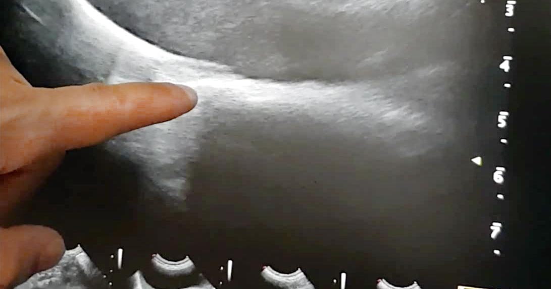
Assessment of the caudal vena cava, via the diaphragmatic hepatic view, is useful for assessing the volume status of a patient.
This is particularly helpful for hypotensive patients, to check whether they require more volume or vasopressor agents, or other management. With regards to “more volume”, this could be crystalloids, colloids or blood products, depending on the patient.
How to find it
- Place the probe under the xiphoid process.
- Increase the depth, adjust the focal point, then reduce the frequency to be able to visualise the diaphragm completely.
- Find the gall bladder, fan laterally to the right (slightly).
- The caudal vena cava is seen deep to the gall bladder as two parallel lines through the diaphragm wall.
- Assessment of volume status – or, more so, fluid responsiveness – is based on interpretation of the degree of collapse during inspiration.
Interpretation
- If it does not collapse during inspiration (“fat”), this can mean the vena cava is adequately loaded or volume overloaded. These patients would most likely benefit from vasopressor agents (or other managements – for example, pericardiocentesis), rather than further volume.
- If it collapses more than 60% (“flat”), the patient requires more volume.
- If it collapses between 20% to 60% (“bounce”), further volume loading can be trialled.

Leave a Reply