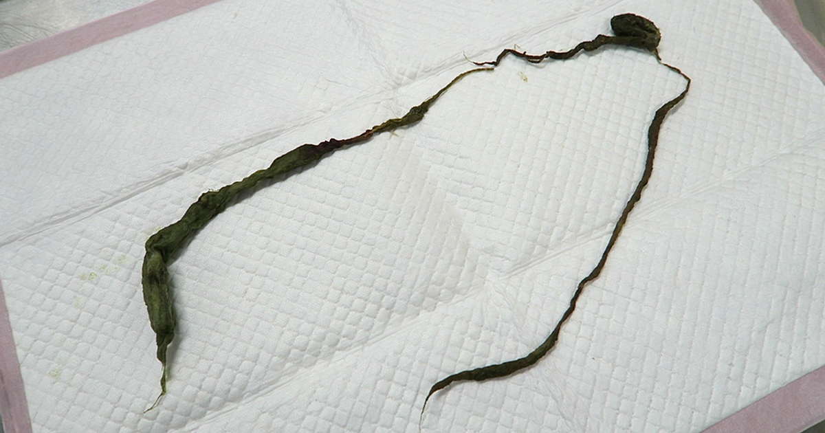
Linear foreign bodies can be tricky to diagnose, compared to normal foreign bodies, for many reasons. Mostly because you often don’t see the classic obstructive pattern appearance on radiographs or ultrasound.
In this short blog series, we are going to cover some hints and tips that can make diagnosing a patient with a linear foreign body easier. Then, we’ll discuss things that should be considered when deciding whether you are the right person to take the patient to surgery…
So, let’s start with radiography.
- Not all patients with a linear foreign body will be completely obstructed. This means you won’t always visualise classic intestinal dilation. In fact, it has been reported that up to 50% of patients with a linear foreign body will not have an obstructive pattern present on radiographs.
- Look for the characteristic small “comma shaped” gas pattern. This is caused by plication and bunching of the small intestine around the tether.
- The small intestine can appear to be bunched up in one area, rather than spaced out around the abdomen. However, obese animals – especially cats – can have “pseudo-bunching” due to large amounts of abdominal fat bunching the intestine together.
- Loss of serosal detail is often seen due to inflammation surrounding the affected intestine.
- Always include a left lateral radiograph in your series. Gastric contents will fall to the fundus on the left of the abdomen and gas will raise to the pylorus, which will highlight the foreign body anchor in the pylorus.
- Perform thoracic radiographs to assess for aspiration pneumonia and a potential oesophageal component of the linear foreign body. If aspiration is present then you know you will need to continue antibiotic therapy postoperatively.
In the next post, we cover some key points for diagnosing linear foreign bodies on ultrasound…

Leave a Reply