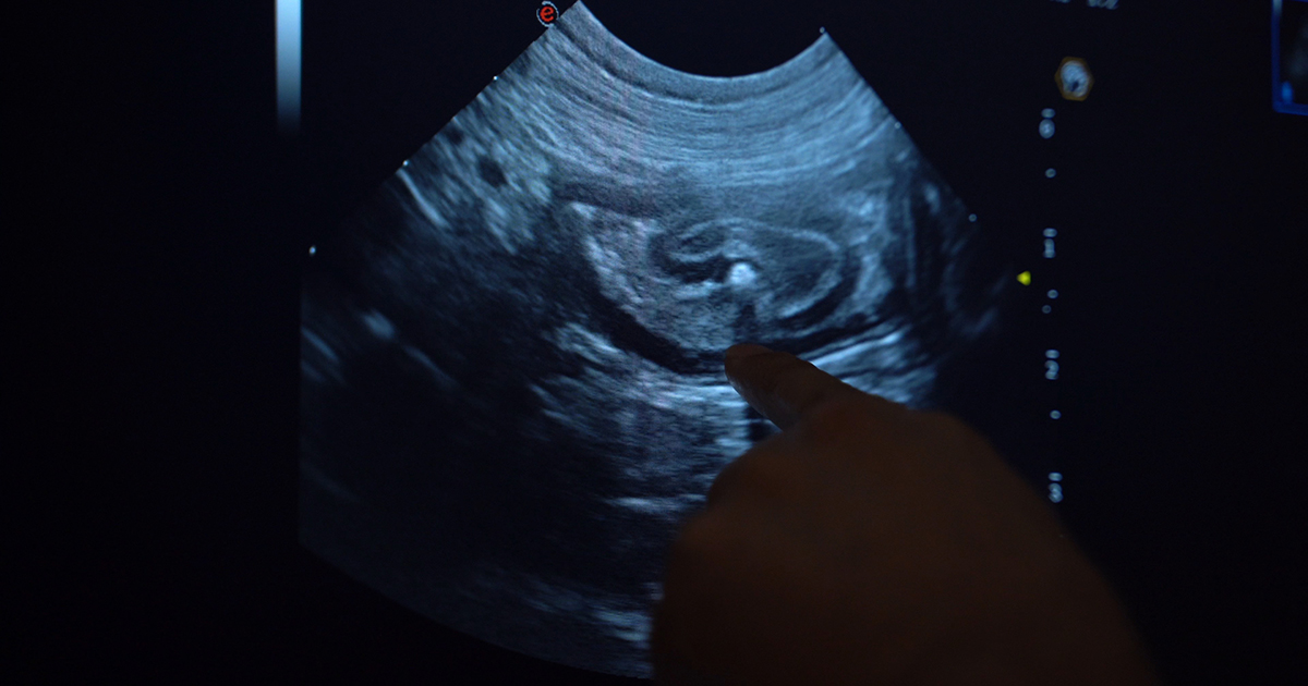
Following on from the previous post where we discussed tips on how to diagnose a linear foreign body on a radiograph, this post sees us cover how to diagnose it on ultrasound.
If used by an experienced ultrasonographer who knows what to look for, ultrasound can be a highly sensitive and specific diagnostic test.
What do we look for?
- Remember not all patients will have intestinal dilation as the linear foreign body may be only causing a partial obstruction. Alternatively, it could be occluding the gastric outflow completely.
- Intestinal plication, which looks like intestinal loops bunching up on each other around the tether.
- A central discrete hyperechoic line running along the middle of the bunching intestine. This bright line is the tether. Often when looking closely enough, the tether will have distal acoustic shadowing as the ultrasound pulses cannot pass through it.
- The leading aboral segment and the trailing adoral anchor will have acoustic shadowing.
- The adjacent mesentery is often hyperechoic compared to other areas in the abdomen, indicating inflammation.
- Gastric dilation with fluid is often seen if the anchor is in the pylorus, as it causes an outflow obstruction.
- Free abdominal fluid may be visible and a sample should be collected for assessment. If bacteria can be demonstrated in one of the following ways:
- By visualising free or intracellular bacteria under the microscope.
- By finding that the glucose is lower (lower than 20mg/dL) and the lactate is higher (2mmol/L) in the abdominal fluid sample compared to peripheral blood then this indicates perforation of the gastrointestinal tract has occurred and septic peritonitis is present.
In the third and final post, we will cover things to consider when deciding whether to perform the exploratory laparotomy yourself, or if you should transfer the patient to a referral facility for surgery.

Leave a Reply