Tag: Anaemia
-

Icteric serum
—
by
The final discolouration of the serum we are going to cover is icteric serum. Icteric serum is caused by the presence of excess bilirubin in the blood stream as a result of increased production (pre-hepatic) or inappropriate excretion (hepatic and post-hepatic). The most common cause of pre-hepatic icterus is haemolytic anaemia, while hepatic disease and…
-

PCV/total solids interpretation: serum colour
—
by
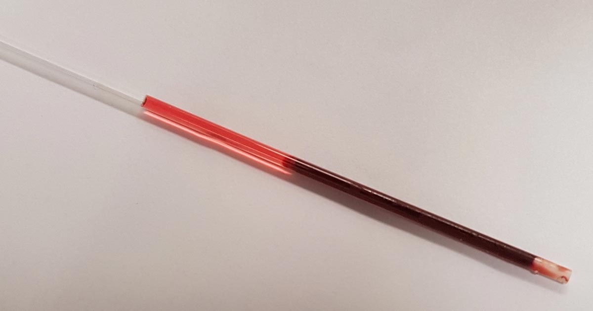
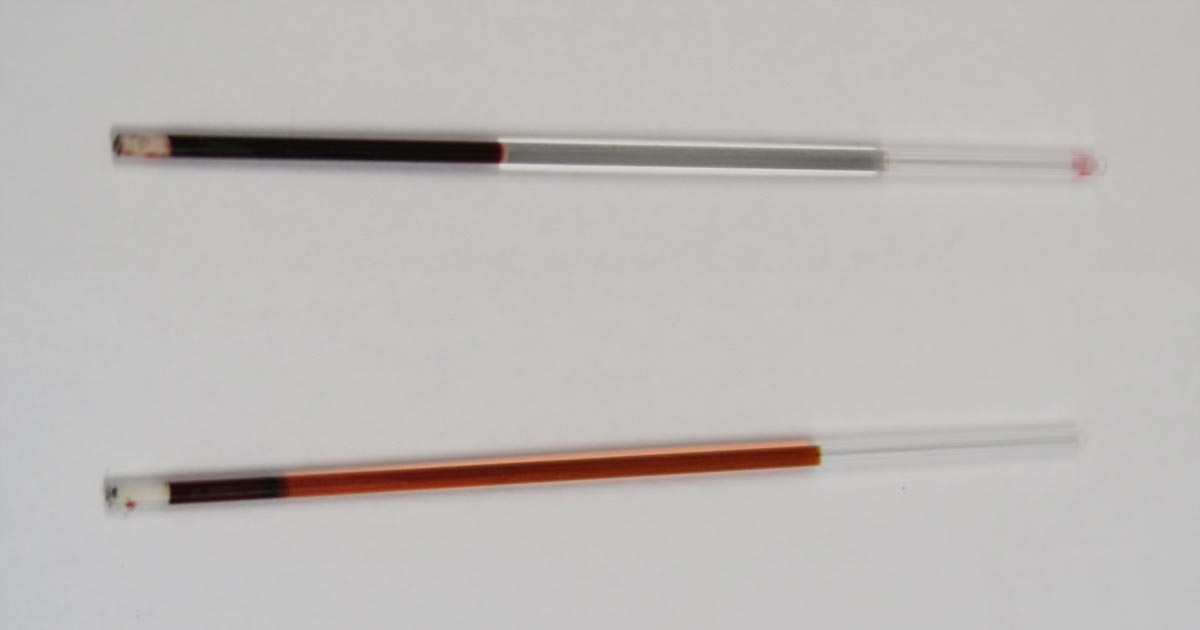
When interpreting the often misinterpreted and underused PCV and total solids test, it is important to take note of the serum colour as this may give clues into the diagnosis. The most common abnormalities seen in clinic are icteric, haemolysed and lipaemic serum. Clear serum can also be of importance – especially when you interpret…
-

PCV/total solids: getting the most from simple test
—
by
The PCV and total solids (TS) test is simple, yet informative – but is often misinterpreted or underused. It is important to remember all test results need to be interpreted in light of the patient’s history, presenting clinical signs and general physical examination findings. The various changes that can be found on a PCV/TS, and…
-

Focus on GDV, part 4: the recovery
—
by
Postoperatively, gastric dilatation-volvulus (GDV) patients remain in our intensive care unit for at least two to three days. Monitoring includes standard general physical examination parameters, invasive arterial blood pressures, ECG, urine output via urinary catheter and pain scoring. I repeat PCV/total protein, lactate, blood gas and activated clotting times (ACT) immediately postoperatively and then every…
-

Focus on GDV, part 1: resuscitation
—
by
Last month we covered a bit of pathophysiology, presenting pathophysiology, presenting clinical signs and the radiographic diagnosis of gastric dilatation-volvulus (GDV). Now we cover the three things you need to do as soon as a suspected case is presented: IV fluid resuscitation decompression of the stomach pain relief Depending on the number of staff you…
-

Christmas dangers
—
by
Christmas can be a busy time for vet clinics, so here is a list of common intoxications and conditions to keep an eye out on during the festive period. Chocolate Numerous online calculators can determine whether a toxic dose has been consumed and they are a great place to start. I always perform emesis in…
-

Pulse oximetry is great, but know its limitations
—
by
Pulse oximetry is a very useful diagnostic and monitoring tool that has become commonplace in veterinary clinics. It measures the percentage of haemoglobin saturated with oxygen, and is an indirect measure of arterial oxygen levels. However, here are several important points to help you understand the limitations of pulse oximetry. Causes for false readings Falsely…
-

Blood smears – make them a routine test
—
by
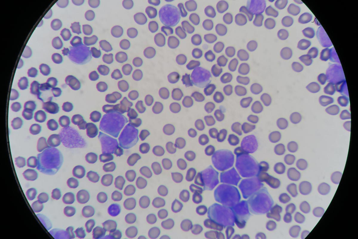
Blood smear evaluation is an often overlooked, but very important, aspect of an in-house haematology. With the advancement in haematology analysers that can now detect reticulocytes and even band neutrophils, some practitioners are beginning to rely solely on the numerical data alone in evaluating the patient’s blood. The art of blood smear interpretation is on…
-

Hormones in practice, part 2: common conditions
—
by
In part one, we broached the taboo of “women’s problems”. A bit of reading about Endometriosis Awareness Month in March and I was staggered about the huge impact our hormonal fluctuations can have on us as individuals, business and the economy. As the editor of Veterinary Woman, it’s my aim to support women in the…
-

AFAST, part 2
—
by
Part one of this series looked at how to perform an abdominal focused assessment with sonography for trauma (AFAST) – this week looks at how to interpret abdominal fluid scores (AFS) in a clinical setting. To recap – the score is out of a possible 4, with each site allocated a 0 or 1 based…