Tag: Clinical signs
-

Ionised hypocalcaemia, pt 3: acute treatment and management
—
by
Treatment of ionised hypocalcaemia (iHCa) is reserved for patients with supportive clinical signs, then divided into acute and chronic management. Since the most common cases of clinical hypocalcaemia in canine and feline patients are acute to peracute cases, this blog will focus on the acute treatment and management of hypocalcaemia. Clinical signs The severity of…
-

Ionised hypocalcaemia, pt 2: eclampsia
—
by
As discussed in part one of this blog series, a myriad of disease processes can lead to ionised hypocalcaemia (iHCa). Despite this, only hypocalcaemia caused by eclampsia and hypoparathyroidism (primary or iatrogenic – post-surgical parathyroidectomy) are severe enough to demand immediate parenteral calcium administration. Hypoparathyroidism is quite rare, so this blog will not explore the…
-
Hyponatraemia, pt 2: causes
—
by
The causes of hyponatraemia can be divided into three major categories, based on serum osmolality. This is further divided based on the patient’s volume status (Table 1). Most patients we see in clinic fall into the hypovolaemic category, except patients with diabetes mellitus. Table 1. Causes of hyponatraemia based on osmolality and volume status (from…
-
Hyponatraemia, pt 1: clinical signs
—
by
Hyponatraemia is a relatively common electrolyte disturbance encountered in critically ill patients, and the most common sodium disturbance of small animals. In most cases, this is caused by an increased retention of free water, as opposed to the loss of sodium in excess of water. Low serum sodium concentration Hyponatraemia is defined as serum concentration…
-
Cardiopulmonary resuscitation, pt 4: monitoring equipment
—
by
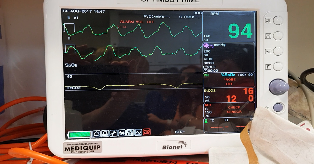
Monitoring devices are crucial in the success of cardiopulmonary resuscitation (CPR). Many monitoring modalities can be used during CPR for the assessment of the adequacy of CPR efforts and the return of spontaneous circulation (ROSC). These monitoring devices, however, should never be used as a sole mechanism to confirm cardiopulmonary arrest. Indeed, commencement of CPR…
-
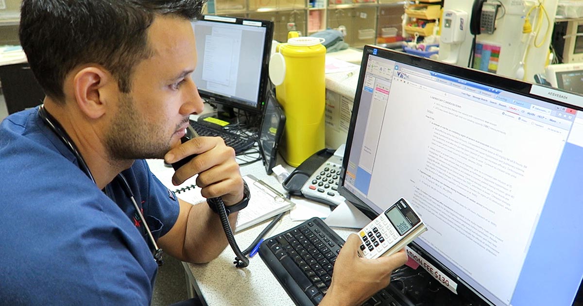
Intoxication: working out possible ingested dose
—
by
We frequently field telephone calls from owners concerned about their pet being intoxicated or having access to a toxic compound. These are the list of questions I always ask owners: What is your pet doing? The main reason I ask this question first is to determine if the pet’s life is in danger. If the…
-
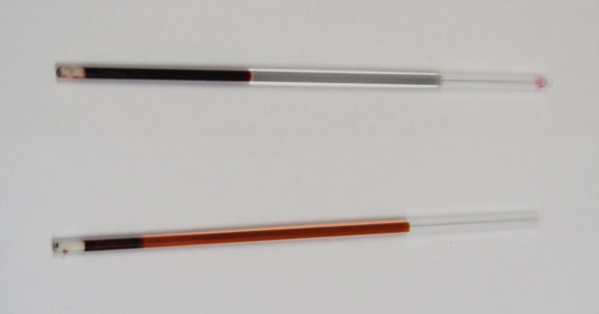
PCV/total solids: getting the most from simple test
—
by
The PCV and total solids (TS) test is simple, yet informative – but is often misinterpreted or underused. It is important to remember all test results need to be interpreted in light of the patient’s history, presenting clinical signs and general physical examination findings. The various changes that can be found on a PCV/TS, and…
-
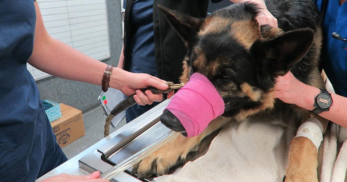
Focus on GDV, part 2: releasing the pressure
—
by
Last week we covered IV fluid resuscitation and pain relief. This week we will go into more detail about gastric decompression. Gastric decompression can be achieved in two ways: trocarisation stomach tube (orogastric tube) placement The decision on which method to use depends on many factors – personal preferences, past experiences and clinical protocols, to…
-
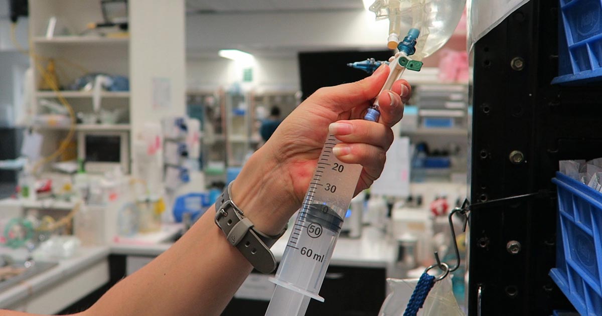
Focus on GDV, part 1: resuscitation
—
by
Last month we covered a bit of pathophysiology, presenting pathophysiology, presenting clinical signs and the radiographic diagnosis of gastric dilatation-volvulus (GDV). Now we cover the three things you need to do as soon as a suspected case is presented: IV fluid resuscitation decompression of the stomach pain relief Depending on the number of staff you…
