Tag: Potassium
-
Blood gas analysis, pt 2: acid-base disturbances
—
by
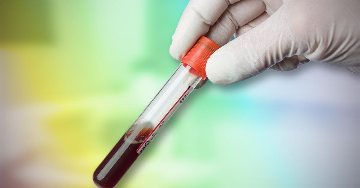
Acid-base disturbances are common in critical patients. These changes must be identified, as even minor deviations from the normal range can lead to significant abnormal body functions. Acidaemia and alkalaemia Acidaemia, which occurs when blood pH falls below 7.35, will lead to: impedance of cardiac output reduced cardiac contractility a blunted response to catecholamine manifesting…
-

Lipaemia – the bane of biochemistry
—
by
Last week we covered haemolysed samples – this week we’re looking at lipaemic samples. Lipaemic samples are caused by an excess of lipoproteins in the blood, creating a milky/turbid appearance that interferes with multiple biochemical tests and can even cause haemolysis of red blood cells. Lipaemia can follow recent ingestion of a meal – especially…
-
PCV/total solids interpretation: serum colour
—
by
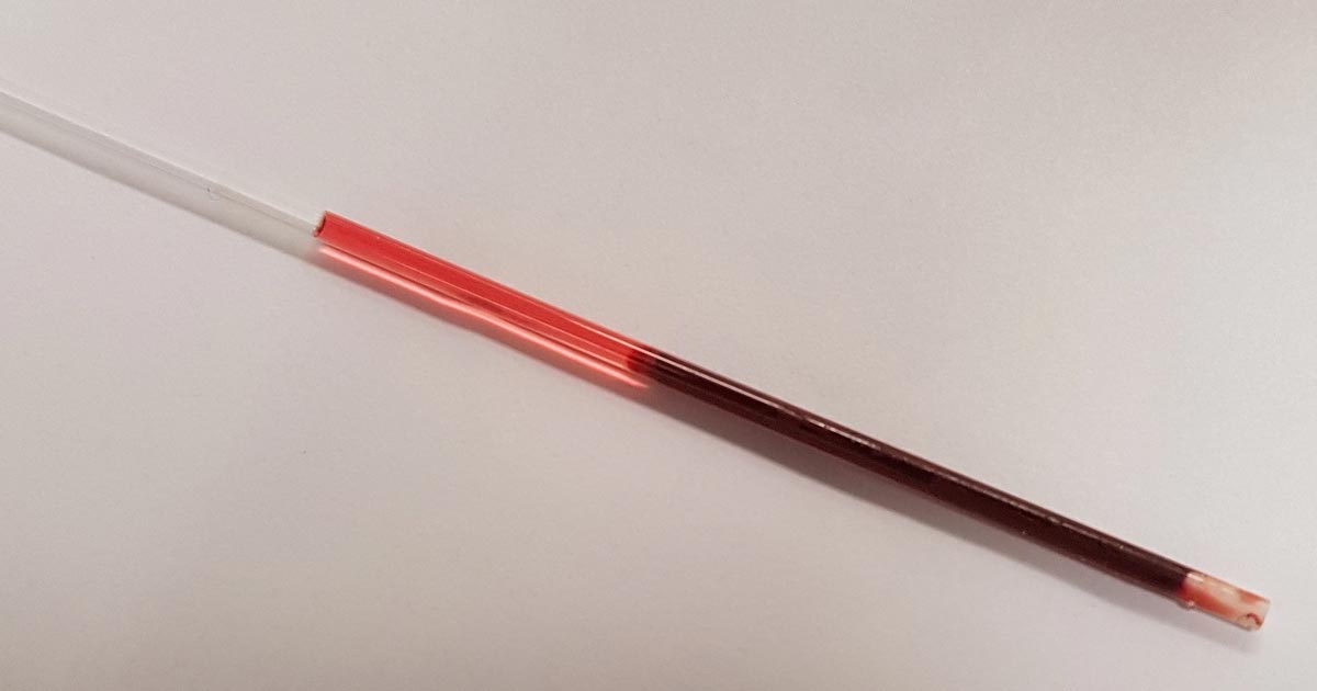
When interpreting the often misinterpreted and underused PCV and total solids test, it is important to take note of the serum colour as this may give clues into the diagnosis. The most common abnormalities seen in clinic are icteric, haemolysed and lipaemic serum. Clear serum can also be of importance – especially when you interpret…
-

Focus on GDV, part 4: the recovery
—
by
Postoperatively, gastric dilatation-volvulus (GDV) patients remain in our intensive care unit for at least two to three days. Monitoring includes standard general physical examination parameters, invasive arterial blood pressures, ECG, urine output via urinary catheter and pain scoring. I repeat PCV/total protein, lactate, blood gas and activated clotting times (ACT) immediately postoperatively and then every…
-
Maintenance fluids
—
by
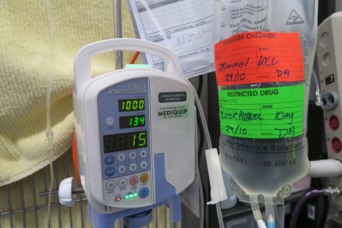
A while ago we discussed the components of a fluid therapy plan and talked about hydration deficits. This week I want to touch on maintenance fluids. Maintenance rates are typically calculated using the following formulae: ml/day = 80 × bodyweight (kg)0.75 (cats) ml/day = 132 × bodyweight (kg)0.75 (dogs) or ml/day = 30 × bodyweight…
-
Making sense of effusions (part 1): is your patient septic?
—
by
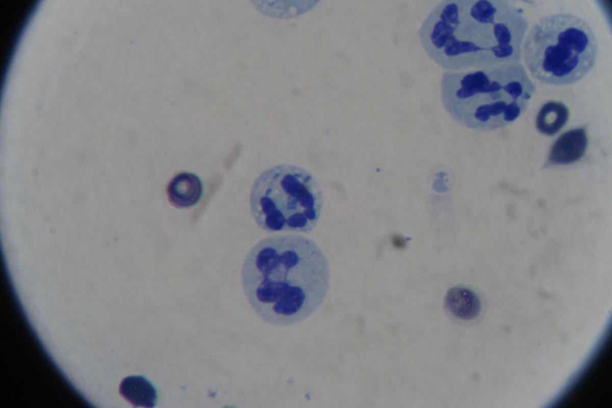
Interpreting effusion samples can be confusing, so try to think of effusions as if you were collecting a blood sample. Many of the in-clinic diagnostic tests that apply to blood samples also apply to effusions, such as: PCV/total protein smears glucose lactate potassium creatinine bilirubin It’s not enough to only check the protein concentration of…
-
SNAP cortisol test
—
by
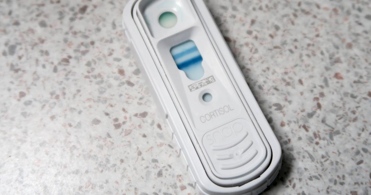
While hyperadrenocorticism is not an uncommon incidental finding in patients presenting to our emergency clinic, hypoadrenocorticism is a lot less common. Or, possibly, more frequently underdiagnosed. Textbook clinical presentations combined with haematology and biochemicial changes can make diagnosis straightforward, but not all patients will present with all the classic signs. To complicate things further, hypoadrenocorticism…

