Tag: Respiratory
-

Laryngeal paralysis
—
by
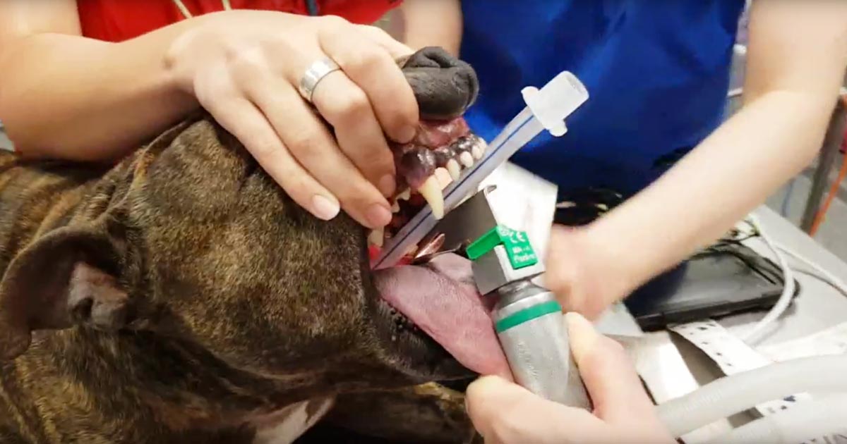
This patient was brought to us for exercise intolerance, breathing difficulty and loud airway sounds. The patient has laryngeal paralysis. This is where the muscles controlling the arytenoids cartilages do not work and leads to failure of opening of the arytenoids during inspiration. Most commonly seen in middle-aged large breed dogs, it can occur acutely,…
-

Blood gas analysis, pt 7: evaluating oxygenation and ventilation
—
by
In patients with respiratory compromise, it is important to look at the respiratory components of the blood gas to determine both oxygenation ability and adequacy of ventilation. To assess oxygenation, the partial pressure of arterial oxygen (PaO2) to fraction of inspired oxygen (FiO2) ratio, and alveolar-arterial gradient (A-a gradient) can be used. Conversely, the partial…
-

Blood gas analysis, pt 2: acid-base disturbances
—
by
Acid-base disturbances are common in critical patients. These changes must be identified, as even minor deviations from the normal range can lead to significant abnormal body functions. Acidaemia and alkalaemia Acidaemia, which occurs when blood pH falls below 7.35, will lead to: impedance of cardiac output reduced cardiac contractility a blunted response to catecholamine manifesting…
-

Blood gas analysis, pt 1: why everyone needs to know about it
—
by
For those of you who have received referral histories from emergency or specialists hospitals, blood gas analysis is probably no stranger to you. For those who have never heard of them before, fear not – you are in for a treat. In my emergency hospital, the blood gas analyser is arguably one of the most…
-

Encouraging mums ‘back to what they love’
—
by
A mother of four appointed as the new head of nursing services at one of the UK’s leading animal hospitals is marking International Women’s Day (today, 8 March) by urging more mums to consider returning to work after having children. Kathryn Latimer Jones has taken on the senior role at Linnaeus-owned Northwest Veterinary Specialists (NWVS)…
-

Focus on GDV, part 4: the recovery
—
by
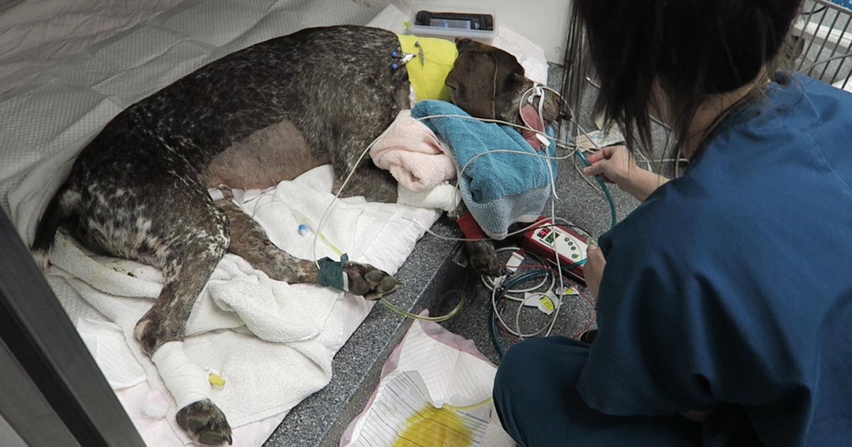
Postoperatively, gastric dilatation-volvulus (GDV) patients remain in our intensive care unit for at least two to three days. Monitoring includes standard general physical examination parameters, invasive arterial blood pressures, ECG, urine output via urinary catheter and pain scoring. I repeat PCV/total protein, lactate, blood gas and activated clotting times (ACT) immediately postoperatively and then every…


